Thanks to a mouse watching clips from 'The Matrix,' scientists have created the largest functional map of a brain to date – a diagram of the wiring connecting 84,000 neurons as they fire off messages.
Using a piece of that mouse’s brain about the size of a poppy seed, the researchers identified those neurons and traced how they communicated via branch-like fibres through a surprising 500 million junctions called synapses.
The massive dataset, published on Wednesday by the journal Nature, marks a step toward unraveling the mystery of how our brains work.
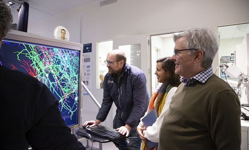
The data, assembled in a 3D reconstruction colored to delineate different brain circuitry, is open to scientists worldwide for additional research – and for the simply curious to take a peek.
"It definitely inspires a sense of awe, just like looking at pictures of the galaxies," said Forrest Collman of the Allen Institute for Brain Science in the United States, one of the project’s leading researchers.
"You get a sense of how complicated you are. We’re looking at one tiny part... of a mouse’s brain and the beauty and complexity that you can see in these actual neurons and the hundreds of millions of connections between them".
How we think, feel, see, talk, and move are due to neurons, or nerve cells, in the brain – how they’re activated and send messages to each other.
Scientists have long known those signals move from one neuron along fibres called axons and dendrites, using synapses to jump to the next neuron.
But there’s less known about the networks of neurons that perform certain tasks and how disruptions of that wiring could play a role in Alzheimer's, autism, or other disorders.
With the new project, a global team of more than 150 researchers mapped neural connections that Collman compares to tangled pieces of spaghetti winding through part of the mouse brain responsible for vision.
How scientists mapped the brain
The first step: show a mouse video snippets of sci-fi movies, sports, animation, and nature.
A team at Baylor College of Medicine in the US did just that, using a mouse engineered with a gene that makes its neurons glow when they’re active.
The researchers used a laser-powered microscope to record how individual cells in the animal’s visual cortex lit up as they processed the images flashing by.
Next, scientists at the Allen Institute analysed that small piece of brain tissue, using a special tool to shave it into more than 25,000 layers and take nearly 100 million high-resolution images using electron microscopes. They then painstakingly reassembled the data in 3D.
Finally, scientists from Princeton University in the US used artificial intelligence (AI) to trace all that wiring and "paint each of the individual wires a different colour so that we can identify them individually," Collman said.
They estimated that microscopic wiring, if laid out, would measure more than 5 km.
Implications for human health
Could this kind of mapping help scientists eventually find treatments for brain diseases?
The researchers call it a foundational step, like how the Human Genome Project that provided the first gene mapping eventually led to gene-based treatments.
Mapping a full mouse brain is one next goal.
"The technologies developed by this project will give us our first chance to really identify some kind of abnormal pattern of connectivity that gives rise to a disorder," said Sebastian Seung, Princeton neuroscientist and computer scientist and another of the project's leading researchers.
The work "marks a major leap forwards and offers an invaluable community resource for future discoveries," wrote Harvard neuroscientists Mariela Petkova and Gregor Schuhknecht, who weren’t involved in the project.
The huge and publicly shared data "will help to unravel the complex neural networks underlying cognition and behaviour," they added.