A robot-guided “smart biopsy” technique has been tested on UK patients for the first time, with researchers hopeful it could spell the end of invasive procedures for those with suspected cancer.
Medics used advanced MRI scans to identify different areas of tumours and take multiple samples at once to better understand their biology.
This could potentially help personalise cancer treatment, they suggest, with hopes that patients could one day forego biopsy completely as doctors would be able to study tumours from scan images the same way they would under a microscope.
By understanding a tumour in more detail, doctors can basically select the treatments that are most likely to work on an individual patient
For the study, led by a team at the Royal Marsden NHS Foundation Trust, 12 patients with suspected etroperitoneal and pelvic sarcomas (RPS) – a rare group of soft tissue tumours that develop in the pelvis and the back of the abdominal cavity – were given smart biopsies.
The MRI scan was performed a week before the biopsy, with sample then taken from three distinct regions of the tumour.
The process is understood to have taken about an hour with patients discharged on the same day.
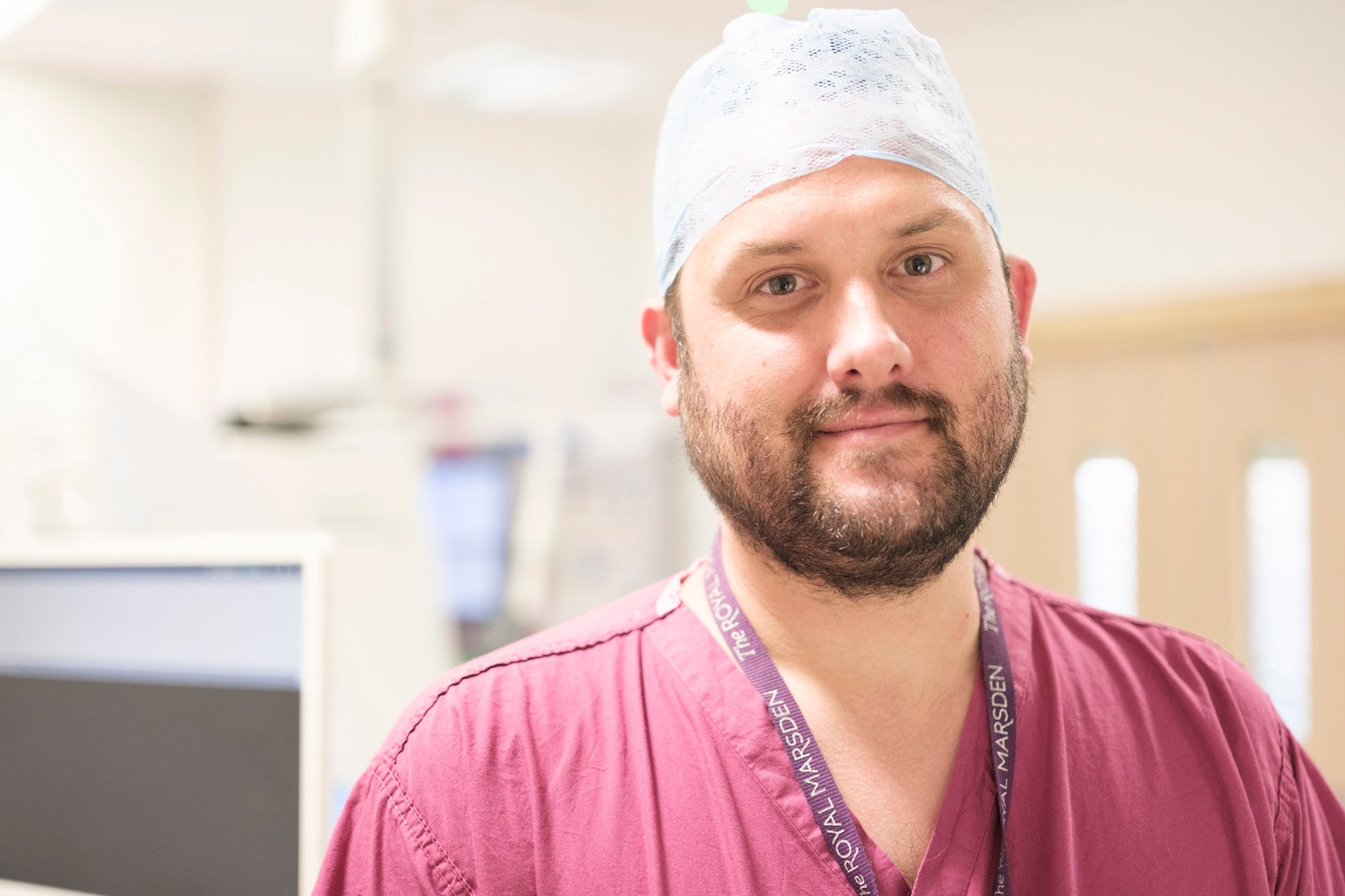
Dr Edward Johnston, consultant interventional radiologist at The Royal Marsden NHS Foundation Trust and The Institute of Cancer Research, London, told the PA news agency: “Current biopsy just involves sampling one region rather than multiple regions. Secondly, it doesn’t have a very detailed MRI acquisition beforehand.
“A lot of this introduces uncontrolled for variation. Biopsy at this point is very similar to how it was in the 1970s really, and a lot of that is actually quite random.
“So we want to make it a lot more thought out.”
Using these scans prior to the robot-guided biopsy allowed researchers to identify different parts of the tumour and sample each area in a planned way.

Dr Johnston said better understanding different areas of a tumour is important for patients as they can behave in different ways and respond differently to treatment.
“Ultimately, it leads to more personalised cancer treatment,” he added.
“By understanding a tumour in more detail, doctors can basically select the treatments that are most likely to work on an individual patient.
“But also in future, if we do more and more of these smart biopsies and understand more and more about imaging signature itself, we really hope that biopsy can be forgotten in some situations.”
Dr Johnston used the analogy of thinking of a tumour “as a fruit bowl”. He said: “You’ve got some apples, some oranges, some bananas.
“And biopsy up until now has basically involved the semi-random sampling of a piece of fruit; it may hit an apple, it may hit an orange.
“And then the assumption there, which is shown to be incorrect in studies up to this point, is that if you hit an orange, then the whole tumour is an orange – that’s not true.”
One day, you could just do an MRI scan, and you would know exactly what that tumour is
He also hopes using advanced MRI scans to look at tumours could eventually allow doctors to assess images in the same way they would under a microscope.
“We ultimately want to look at the relationship between imaging appearances and what is seen under the microscope,” Dr Johnston said.
“So if we validate these imaging appearances versus what’s seen in histology under the microscope, we might one day be able to forego biopsy completely, which would give rise to a digital image-guided biopsy.
“One day, you could just do an MRI scan, and you would know exactly what that tumour is.”
All of the smart biopsies in the study were done within a one hour slot, according to Dr Johnston.
“The robotic technology helped us reduce the time of biopsies a lot,” he told PA.
“They still take a little bit longer than than a than a standard free hand biopsy, but not that much longer.
“To give you a broad idea, everything’s less than an hour. A free hand biopsy takes about half an hour, so it’s not a huge increase.”
The team is now exploring expanding the technique for other tumour types.
“I’m advertising for a PhD student at the moment to do this in myeloma,” Dr Johnson said.
“We have had conversations in about doing it in lymphoma. So basically, we’d like to roll it out into other tumour types.”
Dr Johnston admits the technique is in is early stages and “extremely specialist”, but added: “We do hope that with training and everything it will become easier.”


