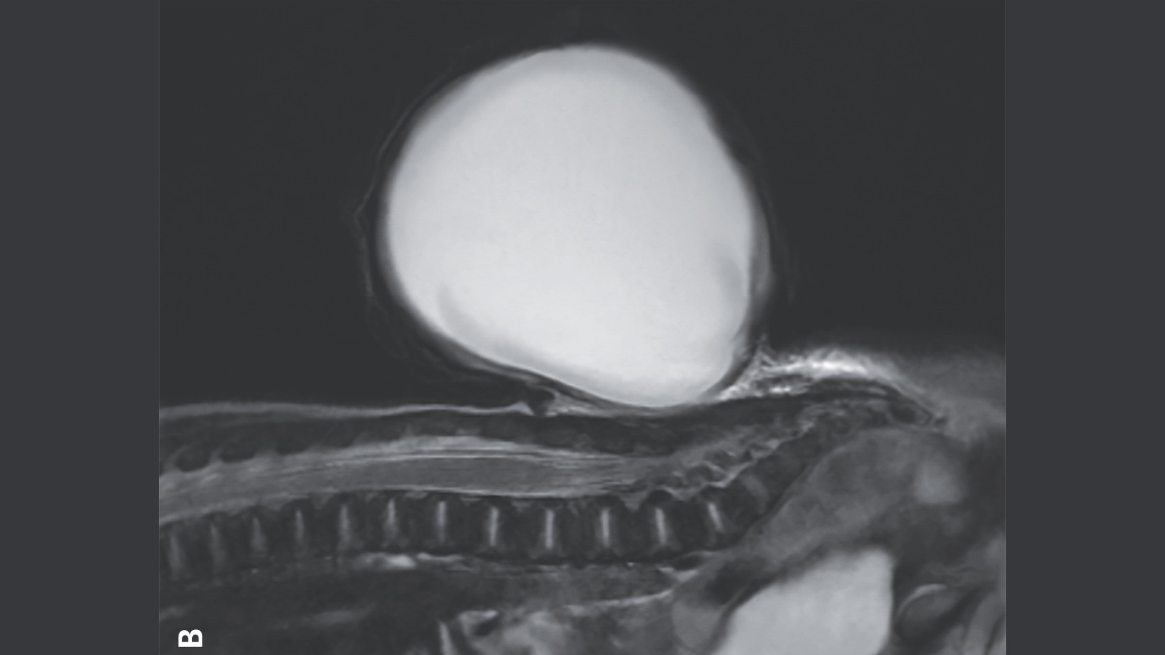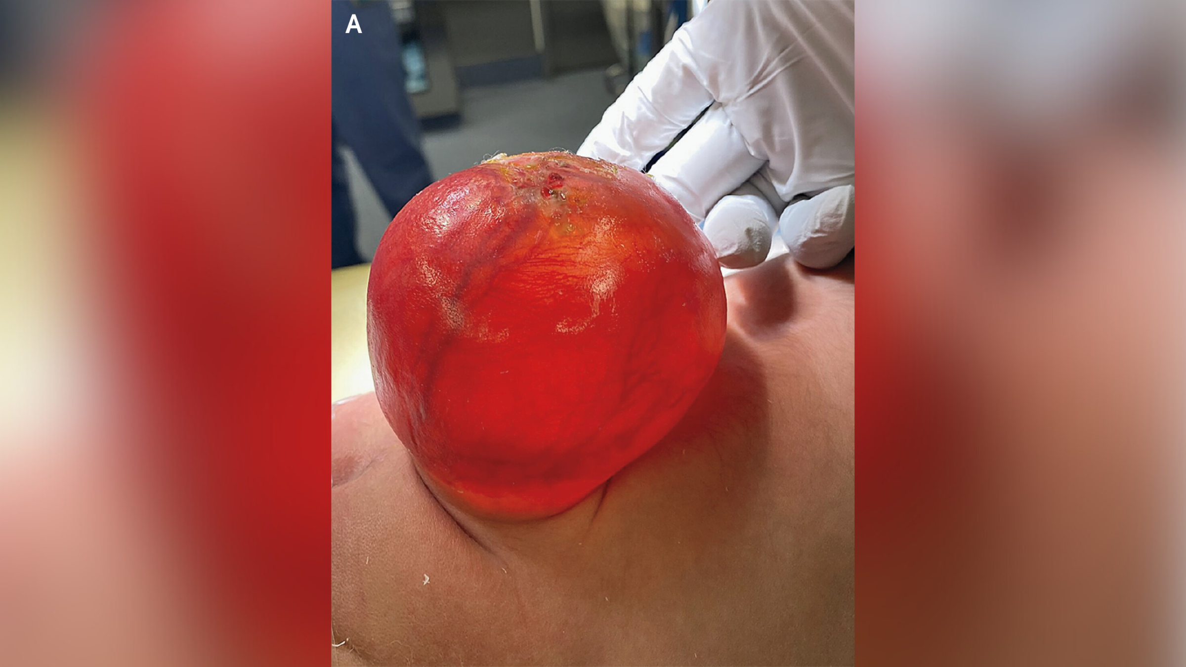
A common birth defect caused a newborn baby to develop a giant, red, balloon-like sac that protruded from the lower back, a striking new image shows.
The image was taken by doctors at Massachusetts General Hospital in Boston. The sac was around 3 inches (7.7 centimeters) long, 2.8 inches (7.1 cm) wide and 2.1 inches (5.3 cm) deep. It was caused by a neural tube defect — which, after heart defects, is the second most common type of disability that is present from birth, affecting between 5 and 8 babies per 10,000 in the U.S.
The neural tube is a hollow structure that forms during the third and fourth weeks post-conception and it later becomes the brain and spinal cord. Sometimes this process is disrupted, leaving babies with a gap in their spine known as spina bifida. Usually, this gap is covered by skin and doesn't cause any symptoms, and many people are unaware they have the condition.
Occasionally, though, tissue and fluid that covers and protects the spinal cord is pushed through the gap, creating a sac-like, protruding structure. This is what happened to the boy in the image, who had a specific version of spina bifida called meningocele.
Related: Mother rejoices after her child's successful spina bifida surgery in the womb
Scientists don't know what causes spina bifida; however a combination of genetic, nutritional and environmental risk factors make it more likely. For instance, if the mother does not have enough folate or vitamin B9 during early pregnancy, takes certain medications such as the anti-epileptic drug valproic acid, or has diabetes that is not well managed, there is a greater risk of them having a baby with spina bifida.
However, none of these factors played a role in the baby's condition in this case, according to a report of his case published Dec. 28, 2024 in the New England Journal of Medicine.


Doctors first noticed the spinal defect during an ultrasound exam around 20 weeks, or halfway through, the pregnancy. Patients with meningoceles often have minor problems such as issues with their bladder and bowels. However, the condition can usually be treated with a simple surgical repair, either before or after birth.
Serious complications are more likely to happen if a baby has another type of spinal bifida known as myelomeningocele, in which neural tissue is also found in the sac. For example, these children may be at risk of developing paralysis, frequent urinary tract infections (UTIs) and a type of brain infection known as meningitis.
In the boy's case, his parents opted for him to have surgery to remove the sac and reconstruct his spinal cord after birth. Four days after the surgery, he was discharged home, and at his six-month checkup, doctors said he was developing without any adverse effects.


