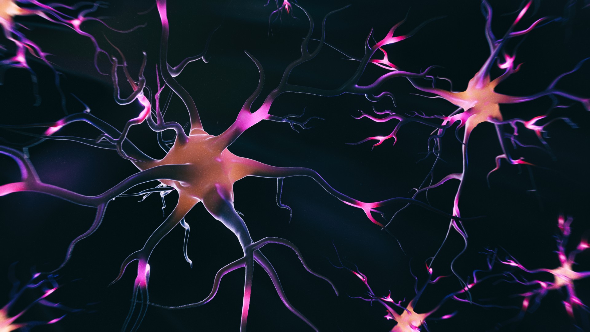
Scientists have provided unprecedented insight into how the activity of neurons in the brain changes across birth.
In a new study, researchers analyzed 184 brain scans collected from 140 fetuses and infants of gestational ages between 25 and 55 weeks post-conception. A typical pregnancy lasts approximately 40 weeks, so these datasets gave the researchers a good snapshot of what the brain looks like before and after birth.
The scans revealed that the activity of neurons within certain regions of the brain increased significantly across birth. These regions include the sensorimotor network, which is responsible for processing external stimuli, like sights and sounds, and for coordinating movements. They also include the subcortical network, which acts like a relay hub for information from different brain regions.
After birth, babies are suddenly exposed to a huge amount of sensory input from the outside world — often, the beeps of hospital equipment, the smells of their parents, and lights shining down on them. Their brains have to be prepared for and able to adapt to this noisy world beyond the womb. However, until now, little was known about how brain activity actually changes across birth, according to the authors of the new study, which was published Nov. 19 in the journal PLOS Biology.
Related: Humans' big brains may not be the reason for difficult childbirth, chimp study suggests
"Birth is the most significant event in human life; it is a really dramatic change from in utero [in the uterus] to the external environment," Lanxin Ji, lead study author and a postdoctoral research fellow at NYU Langone Health, told Live Science. "So there's a lot of change across the whole body, including the brain."
Over a decade, Ji's colleagues built up a detailed dataset of scans of the brains of fetuses and infants using functional magnetic resonance imaging (fMRI). This technique indirectly measures brain activity by monitoring how much oxygenated blood is flowing through the organ and thus is being used by neurons in different areas.
Typically, scientists conduct fMRI by having a person lie very still inside a tube-shaped scanner. However, it is hard to get a clear signal of fetuses' brain activity, especially given that they move around a lot in the womb, Ji said.
To overcome this challenge, the researchers scanned the brains of fetuses using a soft magnetic coil that they put directly onto their mothers' abdomens. Then, they used various analytical techniques, including artificial intelligence (AI), to cancel out the effects of the fetus moving around. With that noise removed, they could reconstruct the neural activity that was unfolding in the fetuses' brains.

Besides the observed effects in the sensorimotor and subcortical networks, the researchers also noticed that brain activity significantly increased in the "superior frontal network" across birth. This network of connected brain areas regulates more complex cognitive skills, such as working memory, which enables people to remember things in the short term — for example, when they're thinking through a math problem or going through a set of directions.
"The change in the superior frontal cortex is kind of beyond our expectation because we believe the frontal lobe develops later during childhood," Ji said. More research is therefore needed to explain these new findings.
Notably, although the researchers observed sharp, significant increases in functional connectivity across birth, the efficiency of communication between these neurons increased much more gradually. In other words, the neurons were connected with others a lot more, but an efficient network had yet to be refined.
The researchers think the brain may need time to refine its network structure to optimize efficiency while removing expendable connections, a phenomenon known as synaptic pruning.
The researchers hope their new findings will act as a scaffold for future research that examines how environmental factors can influence brain development before and after birth. They now plan to compare the timing and growth of brain networks in preterm babies — those born alive before 37 weeks — with those of full-term babies, to see if they differ in their early brain development, Ji said.
Ever wonder why some people build muscle more easily than others or why freckles come out in the sun? Send us your questions about how the human body works to community@livescience.com with the subject line "Health Desk Q," and you may see your question answered on the website!


