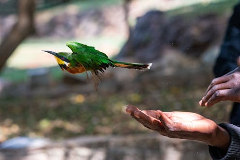
Birds are profoundly important animals. As predators, pollinators, seed dispersers, scavengers and ecosystem bioengineers, the world’s 11,000 species of birds play critical roles in the food chain and therefore the existence of animal life.
They have also shaped the advancement of human societies culturally, philosophically, artistically, economically and scientifically. Birds feature prominently in the history of painting, poetry, commerce and music.
Since they can easily escape from unsuitable habitats, birds are important “sentinel” animals: the number and diversity of species indicates environmental health. BirdLife International’s State of the World’s Birds Report for 2022 says that about half of all bird species are decreasing and more than one in eight of them are at risk of extinction.
Knowledge of bird biology and their place in ecosystems contributes to devising conservation efforts. Biology explains why animals behave the way they do and what threatens their survival.
One of the aspects of bird biology that has long interested scientists is their lungs. They are structurally very complex and functionally efficient. Their lungs are what allows birds to fly. Flying uses a huge amount of energy and some birds fly nonstop over very long distances or at very high altitudes where there is little oxygen.
Even after extensive study, questions about the bioengineering of the avian respiratory system have persisted. They relate to how the airways and blood vessels are shaped, arranged and connected, and how air flows around the lung.
To explore these aspects of the avian lung, my colleagues and I have used a variety of techniques. Three-dimensional (3-D) serial section computer reconstruction is one of them.
Using this technique showed us that the tiny structures (air- and blood capillaries) between which oxygen is exchanged are not the shape they were long thought to be. Because they are so small and so tightly entangled with each other, it wasn’t possible to see their shapes and connections clearly until we used 3-D reconstruction. We were then able to see what makes the bird lung so efficient at taking up the oxygen needed to release energy – key to survival.
The approach
For hundreds of years, scientists could only study biological structures in two dimensions – sections of tissue were placed under a transmission microscope. In the late 1970s, the South African-born Nobel prize winner Sydney Brenner was the first to apply computing to reconstruct series of sections. More recently, 3-D reconstruction methodologies have revolutionised various fields of biology.
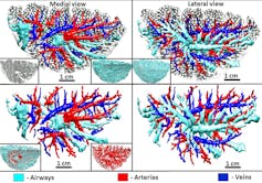
3-D reconstruction showed us that the airways and blood vessels track each other and supply specific parts of the bird’s lung. The various branches of the airway system do not interconnect and neither do the branches of the blood system. We were able to get a much clearer view of the shapes and connections of the air capillaries and blood capillaries in the lung. The compact entwining of the capillaries increases respiratory surface area while minimising the thickness of the blood-gas barrier.
The design of the bird’s lungs forms a highly efficient gas exchange system with large functional reserve. The lungs are ventilated continuously and in one direction (from back to front) with “fresh” air by coordinated actions of the very large air sacs. During every respiratory cycle, the air in the lung is replaced with “clean” air. This maintains a high pressure that drives oxygen into the blood circulating across the lung. It gives birds their flying power.
Our 3-D serial section reconstruction supplied new details and underscored the value of the technique for investigating complex biological structures.
3-D reconstruction
3-D reconstruction entails preparing a spatial model of a structure from 2-D images. Because it takes time, a lot of materials and specialised skills, it’s not often used in biological studies.
We used the method on a chicken lung because this is the model animal for study of the biology of birds.
We cut 2,689 serial sections of a chicken lung at a thickness of 8 micrometres (each micrometre is one millionth of a metre). We stained and mounted them onto glass slides, photographed sections and aligned the images for reconstruction using open-source software.
There are other modern 3-D reconstruction methods that are faster, cheaper and easier to use. But 3-D histological serial section reconstruction (building up a picture from thin slices of tissue) remains a very important technique. The reconstructions have better contrast and signal-to-noise ratio (there’s less unwanted information). Also, dyes and markers can be used to enhance identification of structures.
Bird lung capillaries
The process showed us that the extremely small terminal respiratory units of the bird lung – long called “air capillaries” – are not so: they are rather rotund structures, interconnected by very narrow passages.
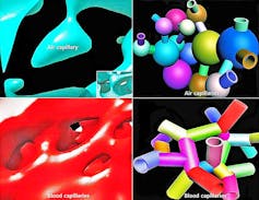
Moreover, the “blood capillaries” are not “true” capillaries like those found in most other tissues and organs that are much longer than they are wide. They comprise clearly separate parts that are about as long as they are wide and interconnect in 3-D. The air- and the blood capillaries of the bird lung intertwine very tightly in a “honeycomb” arrangement.
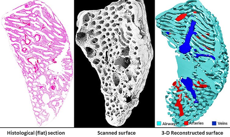
Knowing the shape and size of these units provides information about the gas exchange efficiency of the bird’s lung, which is a flow-through system.
More to come
As more efficient ways of applying 3-D reconstruction technology are developed, 3-D imaging and animation will become a vital means of research in a biologist’s toolbox. It will be possible to fully conceptualise the forms of structural components and hence allow better understanding of how they work.
Vital insights into the biology of animals, including birds, will allow us to formulate more effective measures that will ensure their conservation in the face of challenges from global warming and environmental pollution.
John Maina receives funding from the National Research Foundation of South Africa
This article was originally published on The Conversation. Read the original article.
